Biofilm is an aggregation of bacteria that group together in a structure that enhances their survival and constitutes a significant challenge in treating infections. In addition to bacterial cells, biofilms also contain sugars, nucleic acids, and even proteins. Until recently, it was thought that the protein fibers served only as glue or “scaffolding” within the biofilm structure. A new study has found that fibers composed of these proteins accelerate the degradation of common antibiotic molecules and can protect the entire colony.
Advertisement
Guest post by Dr. Elad Arad, in collaboration with ScienceAbroad
Antibiotic resistance poses a significant obstacle to the treatment of bacterial infections and is an increasingly acute challenge for healthcare systems[1]. Particularly concerning is the formation of clusters, also known as biofilms, by different bacterial species. These clusters adhere to surfaces inside the body and in a patient’s surroundings. They are highly resistant to antibiotic therapy. In addition to bacterial cells, biofilms contain proteins, sugars, and nucleic acids that hold the bacteria within the cluster. These molecules have various functions, such as partially concealing bacterial cells and creating conditions that allow the bacteria to thrive and better withstand antibiotics, thereby prolonging the illness. A new study by Prof. Raz Jelinek’s group at the Ilse Katz Institute for Nanoscale Science and Technology and the Department of Chemistry at Ben-Gurion University of the Negev, in collaboration with researchers from Ben-Gurion University, the Technion, and Aarhus University (Denmark) [2], shows that specific protein fibers in biofilms, namely amyloid proteins, accelerate the degradation of common β-lactam (penicillin-like) antibiotics.
Amyloids are proteins that form strong, sticky fibers under certain conditions. They have gained a bad reputation because amyloid deposits accumulate in the nervous system and in the brain during neurodegenerative diseases, killing nerve cells. Well known examples are Alzheimer’s disease, Parkinson’s disease, and “mad cow” disease. Consequently, newly approved Alzheimer’s treatments aim to inhibit the deposition of these fibers [3]. However, amyloid fibers are widespread and essential in many living systems, including spider silk, silkworm threads, adhesive proteins of marine mollusks and bacterial and fungal biofilms. Their function varies from system to system, and much remains unknown. Previously, it was assumed that amyloids in bacterial systems enable biofilm bacteria to adhere to surfaces, stabilize three-dimensional clusters, and even facilitate the flow of fluids and nutrients into and within the clusters [4].
Recent studies show that some amyloid fibers can catalyze various chemical reactions. For years, researchers have proposed that in the beginning of life chemical reactions were catalyzed not by sophisticated protein enzymes, but by relatively simple protein clusters resembling amyloids. These clusters adsorbed molecules onto their surfaces, making the molecules react more efficiently. These primitive catalytic proteins, the ancestors of modern enzymes, may have functioned this way until the highly efficient enzymes we know today evolved.
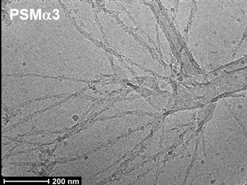
In the present study, the researchers examined various short proteins (peptides) to determine their potential role in degradation of antibiotics. They then investigated the chemical and structural features that promote antibiotic breakdown. Several amyloids typical of Staphylococcus aureus biofilms were investigated, as this pathogen is one of the leading causes of antibiotic-resistant infections [5]. The peptide that was most effective at degrading antibiotics such as penicillin, amoxicillin, and nitrocefin (a cephalosporin) was then identified. Efficiency was assessed in terms of both for the ability to bind and adsorb the antibiotic, as well as for the ability to break it down. Catalytic efficiency was thus assessed by the ratio between degradation rate and binding affinity. These are the two main steps in the interaction between a catalyst (enzyme) and its substrate. Of the S. aureus biofilm components examined, the amyloid peptide PSMα3 proved to be the most efficient relative to its amount.
A central question is whether the structure of amyloid fibers, which are composed of many peptide subunits, drives the accelerated breakdown of antibiotics. This breakdown could undermine the efficacy of the antibiotic treatment. Large natural molecules such as proteins, peptides, and sugars can adopt various spatial conformations, thereby exposing or hiding different parts. The way a protein folds and organizes [6] is critical in determining its activity. Folding dictates which chemical groups are available to react, bind to other molecules, or cleave chemical bonds.
Returning to the study, the researchers approached the question from several angles. They elucidated the fiber structure and used computer modeling to study how the peptides bind to antibiotic molecules within the fiber. They also compared the fiber to derivative amyloids that were designed to have similar yet slightly different structures, which confer distinct chemical properties. Specifically, they examined their capacity to degrade antibiotics efficiently enough to allow bacteria to survive exposure to antibiotics. To confirm that the antibiotics had been degraded, the team used an antibiotic that changes color when broken down. They also tracked structural changes by monitoring molecular weight.
Comparing the properties of various peptides revealed that antibiotic degradation is catalyzed in the presence of peptides that form positively charged amyloid fibers. This is in contrast to other amyloids, which have different charges. These findings highlight the importance of the fibrillar structure and suggest a mechanism involving electrostatic attraction between the positively charged fibers and the negatively charged antibiotics. The researchers demonstrated that adding the native peptide fibers to bacterial growth media, enabled more bacteria to survive exposure to antibiotics. These findings suggest that amyloid fibers in bacterial biofilms serve not only structural purposes, but also provide an additional survival mechanism that helps the bacterial community cope with antibiotics.
The study sheds light on a potential survival strategy employed by bacterial biofilm communities. Understanding these mechanisms could lead to more effective approaches in combating bacterial infections. The study also highlights the role of amyloid protein clusters as catalysts for chemical reactions, paving the way for further research that could mimic these processes to develop new protein-based materials.
Hebrew editing: Smadar Raban
English editing: Gloria Volohonsky
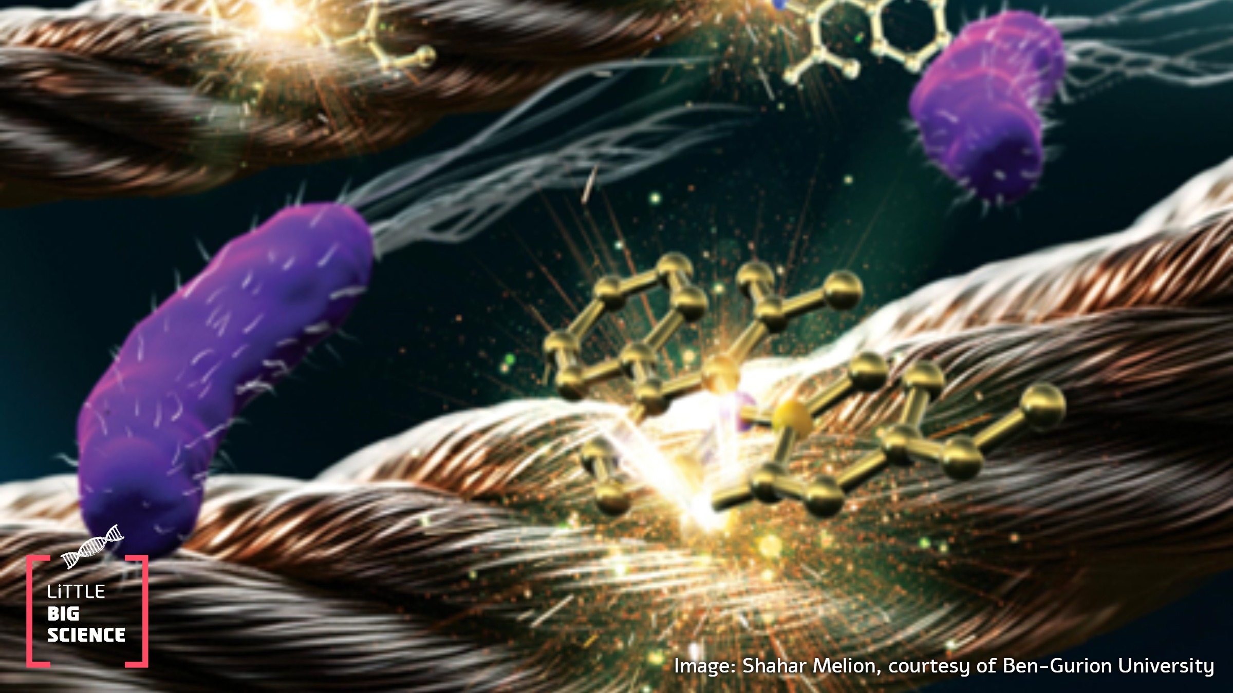
Cover image: bacteria (purple), bacterial protein fibers (bronze), and antibiotic molecules (gold). The active part of β-lactam antibiotics is the square at the molecule’s center (the β-lactam ring). Illustration courtesy of Ben-Gurion University of the Negev. Graphic design: Shahar Melion, Melion Studio.
Dr. Elad Arad completed his studies at Ben-Gurion University of the Negev—a B.Sc. in Chemistry and Philosophy and a Ph.D. in the Department of Chemistry at the Ilse Katz Institute for Nanoscale Science and Technology under the supervision of Prof. Raz Jelinek and Prof. Hanna Rapaport. His doctoral research focused on the surface properties of amyloids, particularly catalytic amyloids. Dr Arad is currently a postdoctoral fellow in the Department of Chemical Engineering at Columbia University, working with Prof. Oleg Gang on the organization and editing of DNA structures to create dynamic assemblies and nanoscale machines.
The non-profit organization ScienceAbroad has been operating since 2006 to maintain ties with Israeli researchers worldwide and facilitate their return to Israel. ScienceAbroad is an international community of over 4,500 Israeli researchers at 300 campuses worldwide. The organization runs 30 centers in North America, Europe, and Australia as well as five online hubs managed by volunteer scientists. It provides tools, builds connections, and opens doors for Israeli scientists wishing to return to Israel, bringing their knowledge, talent, experience, and networks back to academia and industry, serving as a growth engine for the country.
References:
- Post on bacterial antibiotic resistance
- The original research paper upon which this post is based
- Post about the development of an Alzheimer’s drug
- Review article on bacterial amyloids
- “Engineered cells against resistant bacteria” – post on the antibiotic resistance of Staphylococcus aureus
- “Learning to Fold” – post on protein three-dimensional structure


