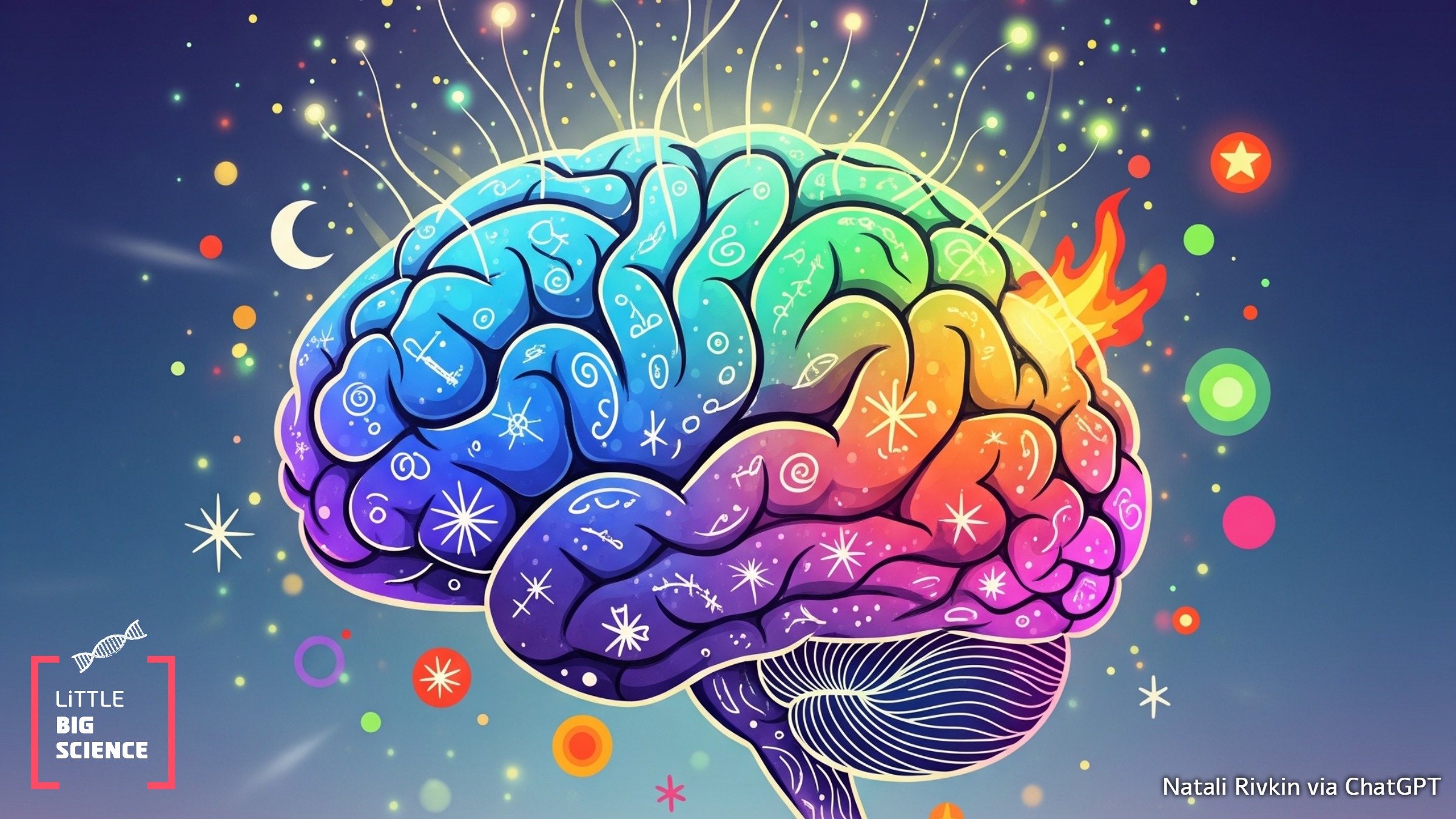
Recent studies reveal that the human brain emits weak photons, aka biophotons, which create a kind of 'aura' around the head. This phenomenon may indicate a role photons play in intercellular communication within the brain.
Advertisement
The human brain is an extremely complex organ. Billions of neurons and other cells, which communicate with each other through a wide variety of electrical and chemical signals, underlie all of our emotions, memories, and thoughts [1]. Despite the tremendous progress science has made in understanding how the brain works at the cellular and molecular level, it can still be regarded as a kind of “black box”: information enters (e.g., via the senses) and information exits (e.g., through bodily movement), yet it is very difficult to study the processes occurring inside the brain—especially if we do not wish to open the subjects’ skulls to peek inside and measure brain activity directly.
For this purpose, non-invasive imaging methods were developed to examine brain activity indirectly from the outside. Two popular techniques are electro־encephalography (EEG) and functional magnetic resonance imaging (fMRI). They exploit phenomena that point to regions where neuronal activity occurs, thereby allowing researchers to identify which areas of the brain are active while a subject performs a defined task. Nevertheless, recent scientific studies reveal a surprising phenomenon: the brain emits a faint flux of light particles, known as biophotons, which can be measured with highly sensitive photon detectors. This light emission, produced as a by-product of metabolic processes such as cellular respiration, adds yet another layer of complexity to brain function.
In a study published in the journal iScience in 2025 [2], scientists used advanced photon detectors to measure the weak stream of light emitted from the human brain. Metabolic processes in brain cells generate photons in the visible-light range of the spectrum, in amounts ranging from single photons to several thousand per square centimeter per second. For comparison, the photon flux from a basic incandescent bulb is about a billion times higher. This phenomenon was also observed in lab-grown neural tissues, indicating that light emission is a fundamental property of active neurons. But what is the link between this cerebral glow and brain activity? Do biophoton emissions from the brain change during different cognitive tasks?
The researchers measured the glow while participants sat in a dark room with their eyes closed, with their eyes open, and while they listened to music, using EEG recordings from the scalp to monitor brain activity. Although the glow shifted as participants moved between tasks, there was no direct correlation between the intensity of this "aura" and the level of electrical activity captured by the EEG. Put simply, the brain does not shine like an LED that brightens when you think hard.
The study also attempted to map biophoton emission from various brain regions, employing focused photon detectors to detect variability among areas. The results indicated that light emission varies across cognitive tasks. For example, the temporal lobe (responsible, among other things, for processing auditory information) displayed one activity pattern when the subjects listened to music and a different pattern when they sat in silence, and this pattern did not match that of the occipital lobe (responsible for visual information processing). However, this variability did not directly match the electrical activity patterns recorded by EEG, suggesting that biophoton emission may reflect metabolic or signaling processes unique to specific areas. These findings raise questions about how local processes in the brain influence the glow and call for further research to map the phenomenon at higher resolution.
The most intriguing part of the study is the hypothesis that biophotons are not merely emitted outward as a by-product of brain activity but may play a functional role in the brain’s intercellular communication network. The researchers believe that much of this light is absorbed within the brain itself and serves as a “messenger” in cell-to-cell communication alongside electrical signals and biochemical processes. This hypothesis was not directly tested in the current study, yet the idea is hardly new. As early as 1923, the scientist Alexander Gurwitsch discovered that onion roots can promote the growth of neighboring roots even when separated by transparent glass; in the presence of an opaque barrier that blocks light, the effect disappeared. This hinted that photons might be involved in intercellular communication [3].
If biophotons do indeed play a role in intercellular communication, the brain may be far more complex than previously thought, integrating optical signaling with electrical and chemical processes. This study is only the beginning, and there is still much to understand about the biophoton phenomenon before we can develop a “photoencephalography” detection technique through which functional brain activity could be inferred. Yet these findings may help us monitor brain activity non-invasively or serve as a new diagnostic tool for detecting cancer or neurological disorders such as epilepsy or autism, in which intercellular communication is disrupted. Who knows—perhaps one day an aura will become a medical diagnostic. Until then, we can take comfort in the fact that even when everything seems dark, our brains are a (very faint) source of light.
Hebrew editing: Shir Rosenblum-Man
English editing: Elee Shimshoni
Sources


